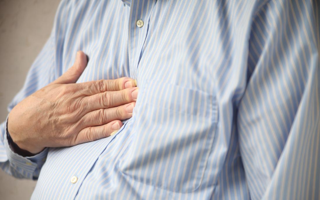
Gastrointestinal causes of non cardiac chest pain
Date:
Wednesday, November 28, 2018
Gastrointestinal causes of non cardiac chest pain
Mohamed El Ghoubary associate proffessor of internal medicine cairo university MD. MRCP •
Gastroesophageal reflux disease
• Esophageal dysmotility
• Peptic ulcer disease
• Biliary disease
• Pancreatitis
• Intra-abdominal masses (benign and malignant) Causes of esophageal chest pain can lead to heartburn symptoms:
• GERD,
• functional heartburn,
• eosinophilic esophagitis,
• achalasia,
• peptic ulcer disease
• or esophageal/
• gastric malignancy. Features suggesting an esophageal as opposed to a cardiac etiology include:
• pain that is nonexertional and prolonged • pain that interrupts sleep • pain that is meal-related • pain relieved with antacids • the presence of additional esophageal symptoms of heartburn, dysphagia, or regurgitation. Diagnosis The typical reflux syndrome can be diagnosed on the basis of the characteristic symptoms without diagnostic testing. Symptom response to antireflux treatment should be used to further cement the diagnosis of GERD prior entertaining any invasive investigation Tools that are currently available for diagnosing GERD include the PPI test, barium esophagram, upper endoscopy, esophageal pH monitoring, and multichannel intraluminal impedance with Ph sensor (MII-pH). The proton pump inhibitor (PPI) test GERD The PPI test is a simple, noninvasive diagnostic tool for GERD that is widely available to community-based physicians . The test is a short course (1–4 weeks) of high-dose PPI given two to three times daily for the diagnosis of GERD. If symptoms disappear or markedly improve with therapy and then return when medication is discontinued, then GERD could be assumed as the diagnosis, and no further testing is required. Barium esophagram Barium esophagram should not serve as the primary test for the evaluation of patients with heartburn. The test is considered positive for GERD diagnosis if reflux is witnessed during examination or if there is morphologic evidence of reflux esophagitis. However, the test has a low sensitivity and specificity for the diagnosis of GERD.
The presence of barium reflux does not necessarily denote GERD, as 20% of normal subjects may demonstrate similar abnormality during esophagram. Barium esophagram may be helpful and should be considered as the first diagnostic tool in patients with GERD who develop dysphagia. In patients with NCCP without alarm symptom, barium esophagram has never been shown to be of value. Upper endoscopy GERD According to most guidelines and consensus statements,upper endoscopy is recommended for GERD patients who do not respond to therapy, those with recurrent or alarm symptoms and to exclude Barrett’s esophagus Upper endoscopy has a low sensitivity for diagnosing GERD, because 50−70% of reflux patients do not demonstrate any evidence of esophageal mucosal injury In the largest study thus far addressing the role of upper endoscopy in NCCP, 44% of the NCCP patients had a normal endoscopy.
Endoscopic findings in those with abnormal endoscopy were GERD-related and included hiatal hernia (28.6%), erosive esophagitis (19.4%), Barrett’s esophagus (4.4%), esophageal stricture or stenosis (3.6%), and peptic ulcer (2%) pH testing pH monitoring use is limited to patients who have not responded to at least a double dose of PPI, patients with normal endoscopy who are candidates for antireflux surgery, and patients who have had antireflux surgery but report recurrence of GERD symptoms Multichannel intraluminal impedance (MII) The MII + pH sensor can determine the nature (liquid, gas, or mixed), proximal extent, and acidity of a reflux event. The technique has been shown to be primarily useful in identifying weakly acid or alkaline reflux in GERD patients who failed PPI twice daily . MII + pH sensor has not been evaluated in NCCP patients, and thus its value in this condition remains unknown. esophageal motility disorders These include spastic (type III) achalasia, diffuse esophageal spasm (DES), and hypercontractile (jackhammer) esophagus.
Peroral endoscopic myotomy Peroral endoscopic myotomy (POEM) was first described in a swine model; 5 years later, more than 2000 of these procedures have been performed in several centers across the world as another treatment modality for achalasia. POEM is classically performed for the treatment of achalasia. However, POEM is potentially an ideal endoscopic therapy for refractory spastic esophageal disorders because it allows myotomy not only of the lower esophageal sphincter (LES) but also of the esophageal body, where the hypertensive contractions occur. Technique of POEM: Briefly, a submucosal bleb is created in the mid-esophagus with saline solution and 0.25% indigo carmine solution. A 2-cm longitudinal mucosal incision was made with a triangular tip knife. The endoscope was then maneuvered into the submucosal space, and the triangular tip knife was used to dissect the submucosal fibers. Subsequently, myotomy of the inner circular muscle bundles is performed, The total length of the myotomy was 19 cm. The mucosal entry was then closed with endoscopic clips.

 info@utopiapharma.com
 Plot No. (2) Industrial Zone (A7) - formerly Zizinia - Cairo - Ismailia Road - 10th of Ramadan - Sharkia